Plasma Wall Interactions
Laboratory
The Laboratory of Accelerators and X-Rays Diffraction has a long standing collaboration with research groups from other research institutions, both national and international. Most of their researchers are members of the Instituto de Plasmas e Fusão Nuclear (IPFN) and this association has been crucial in obtaining funds necessary for its continuous upgrading.
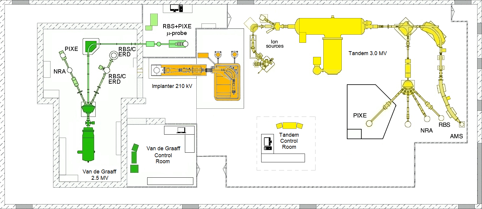
The Ion Beam Laboratory (IBL) hosts two electrostatic accelerators and an implanter. A 2.5 MV Van de Graaff with three experimental beam lines, with all the relevant ion beam techniques available: RBS, PIXE, NRA, Channeling, ERDA and a Nuclear Microprobe. A 3.0 MV Tandem with a micro AMS (Accelerator Mass Spectrometry) line with depth scan profiling capabilities, high resolution PIXE (10-12 eV) setup, a Universal Ion Beam Analysis Endstation and a dedicated line for nuclear physics experiments. A 210 kV Danfysik Ion Implanter with an implantation area of 20x20 cm2 allowing implantations of nearly all the chemical species of the periodic table in the temperature range of 77 K up to 1273 K, in a controlled way. The connection of the implantor to the RBS chamber of the van the Graaff Accelerator, allows situ analysis during the implantation.
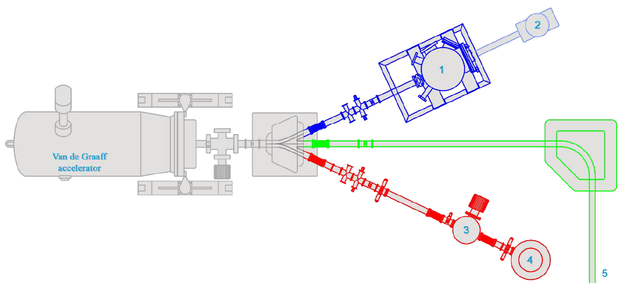
Below is the schematic representation of the different lines associated with the Van de Graaff accelerator: RBS line in red, "Microprobe" in green and the JET line in blue. There are five different experimental stations where the read beam line has two chambers for IBA (3, 4). Both chambers are equipped with surface barrier detectors for RBS, NRA and ERD (only in chamber 4). The chamber 1 on the blue beam line is designed to handle a considerable variety of sample sizes and dedicated to fusion research, where samples, including full JET tiles, contaminated with tritium (T) and beryllium (Be) can be measured. The sample manipulator in this experimental chamber is fully automated and enables analysis over 150 mm along the surface. This allows efficient measurement of whole JET tiles. The RBS particle detector with a 150 μm active detection depth is located at a scattering angle of 150°. The NRA detector is placed at a 135° scattering angle and has an active layer with 2000 μm thickness. A 75 μm thick Mylar stopping foil was selected to allow the detection of D and Be protons from the D(3He,p)4He and 9Be(3He,px)11B reactions whilst stopping the 14 MeV α particles from the 54 9Be(3He,α)8Be reaction. The chamber also allows the X-rays detection for PIXE by means of a "Sirius 30" detector located at a scattering angle of 150o with a Mylar filter 350 μm thick. The chamber 2 is used for PIXE routine analysis. At the end of the green line (5) exists an Oxford Microbeams® type nuclear microprobe with a lateral resolution of 1,5 mm. It is possible to perform PIXE, RBS, STIM (Scanning Transmission Ion Microscopy), CSTIM (Channeled STIM), IBIC (Ion Beam Induced Charge) and IBIL (Ion Beam Induced Luminescence) type of analysis, in normal conditions and external beam.